
For researchers, fast and high-quality analysis of solid tumor tissue samples is important, as is enhancing the quality and reliability of macro-dissection, nucleic acid extraction and molecular profiling. Digital pathology has the potential to empower this research, through automated detection and analysis of tumor cells on H&E-stained tissue sections. With powerful algorithms for research into lung cancer, colorectal cancer and breast cancer, Philips TissueMark is the Philips offering in this space.
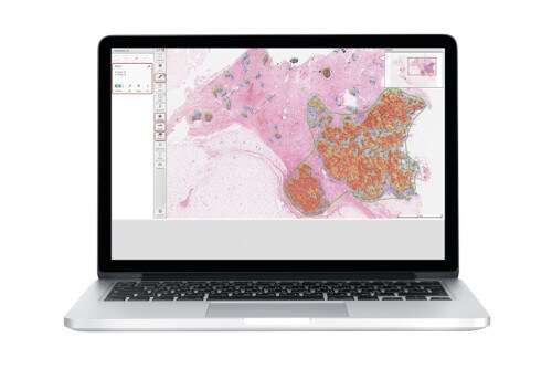
Using automated technology and imaging algorithms, it provides unique methods for tumor cell counting sample quality, sample selection, and tumor sufficiency, even at high sample volumes. Tumor probability is shown via a quantitative visual heat map, based on automatic analysis of tissue samples. The heat map is color-coded and provides information on tumor location and differentiation from stroma, inflammation, and necrosis.
Empowering research studies
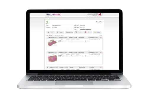
Digital pathology solutions such as Philips TissueMark can be easily embedded within busy molecular research labs to streamline workflow: manage digital slides, approve tissue sample markup, generate results, and manage workflow. It provides a clear and traceable record of tumor regions, tumor burden, and precise boundaries. Key advantages:
Accurate tumor boundaries for macro-dissection
Tumor boundary algorithms automatically annotate the tissue, providing a printout that can be conveniently used for subsequent macro-dissection. All markups are stored indefinitely with the digital slide. Studies have shown1 perfect concordance of molecular test results between manual markup and automated TissueMark annotations for macro-dissection.
Accurate tumor boundaries for macro-dissection.
Automated tumor nuclei counting
The system automatically measures the number of tumor nuclei to provide accurate threshold measurements for percentage tumor nuclei and tumor nucleic acid sufficiency. This helps to ensure the quality and reliability of molecular test results in solid tumor analysis. Detailed studies have shown1 strong correlation between TissueMark calculations and gold standard benchmark data.
Annotated tumor nuclei counting.
Designed to help researchers stay in control
Digital pathology solutions such as Philips TissueMark are designed to support the research workflow. It allows the qualitative analysis of tumor content, enhancing the reliability and quality of molecular profiling. Its powerful and reliable algorithms remove subjectivity from the test, making it easier to establish a solid pattern of consistent and accurate results for tumor burden and tumor boundary annotation across pathology researchers and laboratories.
References
1) Automated tumor analysis for molecular profiling in lung cancer, Hamilton, Peter W. et al, Oncotarget (2015) Vol. 6, No.29
For more information
To learn more, please visit: www.philips.com/digitalpathology